
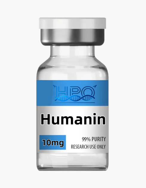
Humanin Peptide: Mitochondrial-Derived Peptide Research Guide
Humanin peptide is a mitochondrial-derived peptide investigated in cellular stress, apoptosis, metabolism and mitochondrial communication. It was first reported in 2001 through a functional screen for factors that protected cultured neuronal cells from selected familial Alzheimer’s disease gene- and amyloid-beta-associated insults. That discovery established a valuable experimental starting point, but it did not establish Humanin as an approved therapy.
For laboratory research only. Not for human or veterinary use. This page describes research questions and analytical controls for a catalog material. Published studies of endogenous Humanin, engineered analogs or specified experimental preparations do not certify the identity, purity, stability, safety or effectiveness of this particular commercial lot.
Identity and specification overview
| Property | Research information |
|---|---|
| Research name | Humanin |
| Class | Mitochondrial-derived peptide (MDP) |
| Genomic context | Short open reading frame associated with mitochondrial 16S rRNA / MT-RNR2 |
| Reference peptide | Frequently described as a 24-amino-acid polypeptide |
| Reference sequence | MAPRGFSCLLLLTSEIDLPVKRRA; verify sequence and terminal state on lot documentation |
| Catalog configuration | Check the selected product variation and vial label |
| Purity and content | Lot-specific analytical results; no universal percentage is asserted here |
| Storage and stability | Follow the current lot label, certificate and validated laboratory procedure |
| Intended use | Qualified laboratory research, not administration |
Humanin is part of a broader group of peptides linked to small open reading frames in mitochondrial ribosomal RNA regions. Humanin and small Humanin-like peptides are associated with MT-RNR2, while MOTS-c is associated with MT-RNR1. This unusual genomic context makes sequence annotation and experimental provenance especially important. Researchers should document whether a paper examined endogenous peptide, synthetic Humanin or a sequence-modified analog.
The 24-residue sequence commonly cited for Humanin is not interchangeable with analogs such as HNG, which contains a residue substitution and may show different experimental activity. The name alone does not describe counterions, terminal modifications, water content or peptide-equivalent content. Confirm these details before comparing mass-based concentrations across lots or publications.
Discovery and evidence boundaries
The original 2001 PNAS study identified Humanin through a functional expression screen and reported protection in selected cultured neuronal-cell models. The effect depended on primary structure and did not extend to every tested cell-death stimulus. That specificity is scientifically useful: it suggests hypotheses about stress pathways, while also showing why Humanin should not be summarized as a universal “cell rescue” factor.
Subsequent literature has examined Humanin in many cellular and animal models. Reviews discuss neurodegeneration, metabolic stress, cardiovascular biology, oxidative stress and apoptosis. These bodies of evidence are heterogeneous. They differ in sequence, model, exposure, comparator and endpoint. Preclinical findings should be presented as model-specific results, not as proof of clinical benefit or longevity extension in people.
Humanin is not represented here as a medicine, supplement, diagnostic marker or anti-aging treatment. No cited reference authorizes personal use of this research material. Claims about disease prevention, memory improvement, insulin sensitivity or lifespan require careful separation of mechanistic observations, animal outcomes and validated human evidence.
Mechanistic research themes
1. Cellular stress and apoptosis
Humanin research frequently examines stress-induced cell death and BCL-2-family signaling. Proposed intracellular interactions include regulation of proteins involved in mitochondrial outer-membrane permeabilization. A protective viability result does not by itself identify the pathway. Researchers should combine viability measurements with pathway-specific endpoints and confirm that assay chemistry is not directly affected by the peptide.
2. Extracellular receptor signaling
Literature also describes extracellular signaling involving receptor complexes and downstream JAK/STAT-related responses. Model selection matters because receptor expression can vary across cell lines, passage numbers and culture conditions. Include receptor-negative or pathway-interference controls when assigning a response to a particular signaling route.
3. Oxidative-stress responses
Humanin has been studied under oxidative, hypoxic and nutrient-stress conditions. Measurements of reactive oxygen species are sensitive to probe chemistry, cell number and timing. A lower fluorescent signal can reflect reduced cell viability or direct interference rather than less oxidative stress. Pair redox readouts with independent measures of viability and mitochondrial function.
4. Mitochondrial communication
As a mitochondrial-derived peptide, Humanin is used to investigate signaling between mitochondria and the rest of the cell. This does not mean that every effect is produced inside mitochondria. Experiments should distinguish extracellular exposure, cellular uptake and intracellular localization instead of inferring location from genomic origin.
5. Metabolic models
Humanin-related studies have examined glucose regulation, insulin action and pancreatic-cell stress in experimental systems. These models can generate testable mechanistic questions, but they do not establish that a research vial treats diabetes or metabolic disease. Record species, tissue, cell state and the exact Humanin or analog sequence used.
Planning a reproducible Humanin experiment
Define the test article: Record the sequence, terminal state, counterion, assigned content and batch. If the study compares native Humanin with an analog, verify both structures rather than relying on catalog names. Use peptide-equivalent content when appropriate and explain every correction factor used in concentration calculations.
Use matched controls: Include vehicle, an appropriate positive control and a negative or sequence control suited to the hypothesis. When the design permits, include a concentration-response series and multiple time points selected before inspecting the outcome. A single concentration and endpoint cannot define potency or mechanism.
Validate the model: Confirm cell identity, passage history and relevant receptor or pathway expression. Distinguish independent biological repeats from multiple readings of the same culture. Randomize sample placement and blind image analysis where feasible to reduce positional and interpretive bias.
Measure more than survival: A viability assay can be influenced by metabolic state and detector chemistry. Combine it with an orthogonal cell-death or membrane-integrity measurement. If mitochondrial function is central to the hypothesis, document the normalization method and ensure that changes are not simply explained by different cell numbers.
Check exposure over time: Nominal starting concentration may differ from the concentration present when the endpoint is measured. Adsorption, degradation and matrix interactions can alter recovery. Analyze samples from relevant points in the experiment and establish stability under the actual conditions used.
Report uncertainty: Provide replicate structure, variability and prespecified exclusions. Retain technically valid null results. Avoid describing an exploratory result as a therapeutic effect, especially when the study uses a cell line, an engineered analog or a model-specific insult.
Analytical quality and documentation
A useful certificate of analysis identifies the lot, test date, chemical form, methods, specifications and results. “High purity” is incomplete without the method and batch. Chromatographic area percentage, peptide mass content and biological activity are different quantities and should not be substituted for one another.
- Identity: Use intact-mass and suitable sequence-supporting evidence to distinguish Humanin from analogs and related peptides.
- Content: Quantify peptide with appropriate standards and state whether the result is on an as-supplied or peptide-equivalent basis.
- Purity: Review separation conditions and related-substance information rather than relying on a percentage alone.
- Water and counterions: Account for non-peptide mass when calculating experimental concentration.
- Application-specific contaminants: Select tests relevant to the planned biological model; chemical purity does not establish sterility or low endotoxin.
- Traceability: Link every working solution, analytical result and experiment to the documented source lot.
Biological activity assays can supplement chemical characterization but cannot replace it. A response may arise from impurities, concentration error or assay interference. Conversely, an inactive result may reflect degradation or an unsuitable model. Investigate discrepancies with orthogonal methods before assigning a mechanistic explanation.
Handling and storage considerations
Follow the current lot label, safety data sheet and institution-approved procedures. Record receipt, opening, storage conditions, container changes and temperature excursions. This page does not assign a fixed 24-month shelf life or a universal solution-storage period because those claims require formulation- and lot-specific stability evidence.
Use a laboratory preparation method appropriate to the intended analysis. Document solvent, pH, concentration basis, container and elapsed time. Verify recovery and stability in the actual matrix. Visual clarity cannot establish chemical identity, biological suitability or absence of contamination. This page intentionally provides no personal dosing, injection or treatment instructions.
Frequently asked questions
1. What is Humanin peptide?
Humanin is a mitochondrial-derived peptide associated with a short open reading frame in the mitochondrial 16S rRNA region. It is commonly described as a 24-amino-acid peptide and is studied in cellular-stress and survival models.
2. Is Humanin an approved anti-aging or neuroprotective treatment?
No approval or treatment claim is made. Research includes cell and animal models, but those findings do not establish clinical safety or effectiveness for this catalog material.
3. Is Humanin the same as HNG?
No. HNG is a sequence-modified Humanin analog often used in research and may differ in experimental activity. Researchers should verify the exact sequence rather than grouping analog data under one name.
4. Does mitochondrial origin prove an intracellular mechanism?
No. Humanin literature discusses both intracellular interactions and extracellular signaling. Experiments need localization and pathway controls before assigning where or how a response occurred.
5. Can a purity percentage establish suitability for cell assays?
No. Purity, content, identity and application-specific contaminants address different questions. Review the methods and choose additional tests appropriate to the biological model.
6. What storage period should a laboratory use?
Use current lot documentation and stability evidence for the actual formulation and matrix. Do not borrow a shelf life from another peptide, analog or supplier without verification.
7. Does this page provide an animal or human dose?
No. The product is restricted to laboratory research. Experimental conditions must follow authorized institutional protocols and appropriate material characterization; vial content is not an administration recommendation.
Related research materials
These links support comparison of distinct research contexts; they are not recommendations for combining products or administering them.
- MOTS-c — a distinct mitochondrial-derived peptide linked to MT-RNR1.
- SS-31 — mitochondrial membrane and cardiolipin research.
- Epithalon — a separate short-peptide research literature.
- Semax — distinct neurobiological research material.
- Selank — separate peptide identity and signaling context.
- FOXO4-DRI — a different cellular-stress research tool.
- ARA-290 — tissue-protective receptor research context.
- GHK-Cu — a distinct copper-binding tripeptide.
Scientific references
- Original Humanin discovery and neuronal-cell rescue study, 2001.
- Humanin as a mitochondrial-derived peptide, 2013.
- Humanin and stress-resistance mechanisms, 2013.
- Humanin in cancer and degenerative-disease research, 2018.
- Humanin and diabetes: review of in vitro and in vivo studies, 2022.
- Evolutionary analysis of Humanin and related mitochondrial peptides, 2023.
Research-use statement: Humanin is supplied for laboratory research only. It is not intended for human or veterinary consumption, diagnosis, treatment or cosmetic application. Qualified researchers are responsible for lawful procurement, institutional authorization, safe handling and appropriate disposal.
Reviews
There are no reviews yet.