
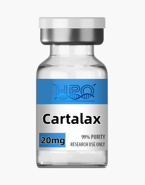
Cartalax
$150.00
Cartalax is the Ala-Glu-Asp (AED) tripeptide used in laboratory studies of chondrocytes, cartilage-matrix markers and chondrogenic differentiation. Research use only.
DISCLAIMER:
Cartalax Peptide Research Guide: AED Tripeptide and Cartilage Models
Cartalax peptide is the short linear tripeptide Ala-Glu-Asp, commonly abbreviated AED. It is studied as a sequence-defined reagent in chondrocyte, mesenchymal stem-cell and cartilage-aging models. Its compact structure makes it useful for investigating how a three-residue peptide may influence cell phenotype, extracellular-matrix markers and stress-associated signaling under controlled laboratory conditions.
The evidence base is small. Most Cartalax-specific reports come from cell-culture work and a limited group of reviews; they do not establish a clinically validated treatment for osteoarthritis, cartilage injury or any other condition. Results should therefore be described as experimental observations, not as proven human benefits. This product is supplied strictly for laboratory research and is not intended for human or veterinary use.
Chemical identity and reference properties
| Property | Research reference |
|---|---|
| Common name | Cartalax (also rendered Kartalax in some literature) |
| Sequence | Ala-Glu-Asp (AED) |
| Systematic description | Alanyl-glutamyl-aspartic acid |
| PubChem CID | 87815447 |
| Molecular formula | C12H19N3O8 |
| Calculated molecular weight | 333.30 g/mol |
| Structure class | Linear acidic tripeptide |
| Typical catalog form | Lyophilized research material |
| Quality basis | Review the certificate of analysis for the supplied lot |
| Intended use | Laboratory research only |
Alanine provides a small nonpolar side chain, while glutamic acid and aspartic acid each add an acidic carboxyl group. Consequently, pH and ionic strength can affect net charge, solubility, chromatographic retention and interactions with matrix components. Researchers should record buffer identity, pH, salts, temperature and peptide concentration whenever comparing results across experiments.
Sequence order is part of identity. Ala-Glu-Asp is not interchangeable with Glu-Ala-Asp, Ala-Asp-Glu or the four-residue peptide Ala-Glu-Asp-Gly. These compounds have different molecular masses and potentially different behavior. Identity testing should distinguish the intended sequence from free amino acids, deletion products, positional isomers and synthesis-related impurities.
Current Cartalax research evidence
Chondrocyte senescence-associated phenotype
A 2023 report compared AED peptide with a cartilage polypeptide complex in cultured chondrocytes. The authors characterized a senescence-associated secretory phenotype using markers that included p16, p21, p53, TNF-alpha, IL-1alpha and SIRT1. They reported that AED exposure changed the measured pattern toward the non-senescent comparison. This supports replication in carefully defined chondrocyte systems, but it does not prove reversal of organismal aging or repair of human cartilage.
Chondrogenic differentiation markers
Another 2023 study examined human mesenchymal stem cells undergoing replicative aging. At the concentration tested in that paper, AED was associated with increased gene expression and protein synthesis of SOX9, aggrecan, type II collagen and COMP. These are relevant markers of chondrogenic differentiation, yet marker expression is not equivalent to production of durable, mechanically functional articular cartilage. Independent studies should add matrix composition, biomechanical testing and longer-term phenotype assessment.
Osteoarthritis-oriented review literature
A review of chondrocyte aging and osteoarthritis discussed AED/Kartalax alongside a cartilage-derived polypeptide complex. The review proposed that effects on pro-apoptotic, proliferative and inflammatory components of the senescence-associated secretory phenotype could be relevant to peptide chondroprotection research. This remains a research framework. The available literature is not sufficient to establish a disease-modifying osteoarthritis therapy or a validated human dosing regimen.
Limits that matter
Cartalax-specific publications are few, study groups are concentrated, and some articles are available principally as abstracts in English. There is no broadly accepted molecular receptor for AED, and the existing reports do not establish pharmacokinetics, biodistribution, long-term safety or clinical efficacy. A responsible interpretation separates direct measurements from proposed mechanisms and avoids converting in vitro changes into medical claims.
Research applications
1. Chondrocyte phenotype studies
Primary chondrocytes or validated cell lines can be used to test whether AED alters viability, proliferation, morphology or expression of cartilage-related genes. Useful endpoints include SOX9, COL2A1, ACAN and COMP, together with hypertrophic or dedifferentiation markers such as COL10A1 and RUNX2. Results should be normalized to cell number and confirmed at both RNA and protein levels where possible.
2. Extracellular-matrix balance
Cartilage homeostasis depends on the balance between matrix synthesis and degradation. Experimental panels may include type II collagen, aggrecan, sulfated glycosaminoglycans, MMP3, MMP13, ADAMTS4 and ADAMTS5. Measuring secreted matrix fragments as well as intracellular expression helps determine whether a marker change corresponds to altered matrix turnover.
3. Cellular-senescence models
Replicative aging, oxidative stress or cytokine exposure can create different senescence-like states. Researchers can measure p16, p21, p53, SIRT1, senescence-associated beta-galactosidase, DNA-damage foci and secreted cytokines. Because no single marker defines senescence, a multi-parameter panel and an appropriate positive control are essential.
4. Inflammatory challenge models
IL-1beta or TNF-alpha challenge is often used to model selected inflammatory aspects of osteoarthritis in vitro. AED may be evaluated for effects on cytokine output, NF-kappaB-related readouts, matrix-degrading enzymes and cell viability. These simplified systems cannot reproduce the entire joint, so conclusions should remain specific to the tested cells, stimulus and exposure window.
5. Mesenchymal stem-cell differentiation
Human mesenchymal stem cells allow investigation of lineage commitment and matrix formation. A rigorous design should include a validated chondrogenic medium, vehicle control, time-course sampling and donor-level biological replication. Pellet size, histology, glycosaminoglycan content and mechanical properties provide stronger evidence than gene expression alone.
6. Structure-activity and stability studies
Useful comparators include reversed or scrambled sequences, constituent amino acids, D-amino-acid analogs and terminally modified variants. LC-MS can track intact AED and potential hydrolysis products over time. These experiments help determine whether any response requires the intact Ala-Glu-Asp sequence or may instead reflect nonspecific nutrient, pH or ionic effects.
Recommended experimental controls
- Vehicle control: match solvent, salts, pH and handling conditions.
- Positive control: verify that the chosen chondrogenic, inflammatory or senescence assay can respond.
- Sequence control: use a reversed or unrelated tripeptide to test sequence specificity.
- Constituent control: compare an equimolar alanine, glutamate and aspartate mixture.
- Viability control: distinguish pathway modulation from cytotoxicity or metabolic suppression.
- Orthogonal validation: confirm key results using an independent analytical method.
Use biological replicates from independent preparations or donors rather than relying only on repeated wells. Randomize sample order when practical, blind image analysis, prespecify exclusion rules and correct for multiple comparisons. Report the exact lot, concentration basis, exposure time, passage number, cell density and statistical model.
Lot-specific quality review
Generic product text cannot establish the properties of every lot. Before quantitative work, review the lot certificate of analysis and distinguish HPLC area purity from net peptide content and gross vial mass. Depending on the study, useful documentation may include:
- identity by mass spectrometry and chromatographic retention;
- purity by a stated HPLC or UHPLC method;
- water, counterion and residual-solvent information;
- net peptide content or quantitative assay value;
- related substances and sequence-isomer assessment;
- batch number, storage conditions and traceability;
- endotoxin, bioburden or sterility only when specifically tested.
For concentration calculations, use the mass basis documented for the lot. Water and counterions may contribute to powder mass, so assuming that total vial mass equals pure AED can introduce systematic error. Analytical recovery should also be checked because small polar peptides can interact with containers or behave differently across chromatography systems.
Handling and storage principles
Follow the product label and lot documentation. Keep lyophilized material sealed, dry and protected from unnecessary light and temperature cycling. Allow a cold sealed vial to equilibrate before opening to reduce condensation. Select a solvent or buffer that is compatible with the intended analytical or cell assay, and document pH and ionic strength.
Prepare solutions using calibrated equipment and an aseptic technique appropriate to the experiment. Minimize repeated freeze-thaw cycles by using validated aliquots when suitable. Establish stability under the actual working conditions with a stability-indicating analytical method; do not assume a universal solution shelf life. This page intentionally provides no self-administration, injection or clinical dosing instructions.
Frequently asked questions
What is Cartalax peptide?
Cartalax is a sequence-defined linear tripeptide composed of alanine, glutamic acid and aspartic acid in that order. It is abbreviated AED and is studied mainly in chondrocyte, cartilage and chondrogenic-differentiation models.
Is Cartalax proven to repair cartilage or treat osteoarthritis?
No. Limited cell-culture reports describe changes in selected differentiation, matrix and senescence-associated markers. Those observations do not establish structural cartilage repair, clinical efficacy or a disease-modifying osteoarthritis treatment in humans.
What mechanism has been established?
Published work suggests effects on panels that include SOX9, aggrecan, type II collagen, COMP, p16, p21, p53, inflammatory cytokines and SIRT1. A single validated receptor or complete molecular pathway has not been established, so mechanism claims should remain hypothesis-driven.
What concentration should researchers use?
There is no universal concentration. Select a range based on the model, solubility, viability and the specific literature being replicated. Include concentration-response testing and report whether values are based on gross powder, net peptide content or another lot-specific assay.
Which controls are most informative?
Vehicle, positive, reversed-sequence or unrelated-peptide, constituent-amino-acid and viability controls are useful. Confirm important observations with orthogonal assays and independent biological replicates.
How should Cartalax be stored?
Use the conditions stated on the label and lot documentation. Keep the dry material sealed and avoid moisture and repeated temperature cycling. Validate solution stability in the exact buffer, container and temperature used by the study.
Is Cartalax intended for human or veterinary use?
No. This product is a laboratory research material only. It is not supplied for consumption, injection, diagnosis, treatment, disease prevention, veterinary use, cosmetic application or household use.
Related research materials
Comparative programs may also review BPC-157, TB500, GHK-Cu, KPV, Vilon, Thymalin, Epithalon and Pinealon. Each material has a different sequence and evidence base and should not be treated as a direct substitute for AED.
Selected references
- PubChem. Alanyl-glutamyl-aspartic acid, CID 87815447.
- Peptides prevent the formation of the aging-associated secretory phenotype of chondrocytes.
- Influence of AED peptide on chondrogenic differentiation of human mesenchymal stem cells during replicative aging.
- Chondrocyte secretory phenotype, osteoarthritis and peptide bioregulation review.
- Chondroinductive peptides for cartilage regeneration: evidence and limitations.
Research use only. Not for human or veterinary use, diagnosis, treatment, prevention, food, cosmetic or household applications. Researchers are responsible for institutional review, risk assessment and compliance with applicable laws and policies.
| Specification | 20mg*10vials |
|---|
Only logged in customers who have purchased this product may leave a review.
Reviews
There are no reviews yet.